Fibroadenoma is a benign organ-specific tumor of the mammary gland of glandular origin. A characteristic difference between fibroadenoma and adenoma is the predominance of connective tissue of the stroma over glandular parenchyma.
The causes of fibroadenoma of the mammary gland are not precisely understood.
The peak of detection occurs at 20-30 years. At the same time, asymptomatic fibroadenoma can be first detected at a much later age during a preventive examination or accidentally during palpation if it is located superficially.
Most often, fibroadenoma is detected as a single tumor of the mammary gland, but there are often cases of multiple fibroadenomas, which can be localized simultaneously in both mammary glands.
Clinical picture of fibroadenoma
Fibroadenoma has a characteristic clinical picture:
- During examination, a large fibroadenoma can be determined visually.

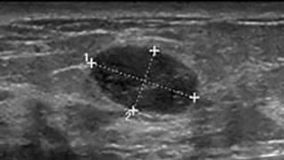
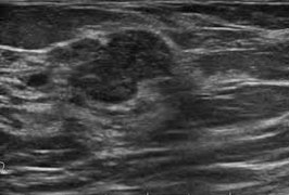
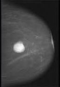
- On palpation it is defined as a clearly demarcated, displaceable tumor of dense-elastic consistency from 1 to 5 cm in largest diameter. [image]
- It is usually located outside the areolar zone. The most common localization is the upper outer quadrant of the mammary gland.
Diagnostics and differential diagnostics
To establish an accurate diagnosis, in the vast majority of cases the following methods are sufficient:
- Clinical examination and palpation of the mammary gland
- Ultrasound of the mammary gland
- Fine-needle aspiration of the tumor with subsequent cytological examination
Differential diagnostics should be carried out with the following diseases:
- Breast cancer
- Breast cyst
- Cystaden papilloma
Treatment and medical tactics
The doctor's tactics in treating fibroadenoma are determined by two main properties of fibroadenoma:
- Fibroadenoma rarely responds to conservative treatment.
- Fibroadenomas are not capable of malignancy/malignancy (except for leaf-shaped fibroadenoma, which in 10% of cases can degenerate into breast sarcoma)
Based on these two facts, the indications for surgical treatment of fibroadenoma of the breast are:
- Leaf-shaped structure of fibroadenoma (absolute indication)
- Large sizes (more than 2 cm) or sizes causing a cosmetic defect.
- The patient's desire to remove the tumor and forget about it.
- Rapid tumor growth.

In other cases, after morphological confirmation of the diagnosis, fibroadenoma can be observed. For surgical treatment of fibroadenoma, we choose the method of tumor enucleation through a paraareolar "cosmetic" incision, usually under local anesthesia, sometimes under general anesthesia.
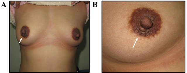
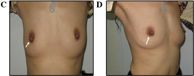
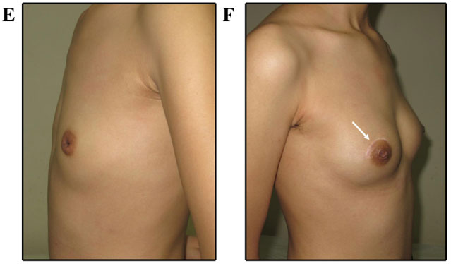
Photographs (A and B) 2 years after fibroadenoma removal. (C and D) photographs 6 months after breast lobe removal. Patient photographs (E and F) 6 months after fibroadenoma removal.
Prevention of fibroadenoma
There are no specific methods of primary prevention. For secondary prevention, regular examinations are carried out using ultrasound of the mammary gland and self-examination once a month from the 5th to the 12th day of the menstrual cycle.

Sometimes new fibroadenomas appear at the site of a removed fibroadenoma or in another place in the mammary gland. It is important to understand that this is always a new fibroadenoma, and not a relapse of the old one.

